Cardiac
Investigations
Vascular
Investigations
Programs
Cardiac Investigations
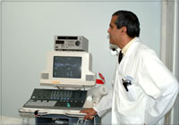
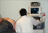
Echocardiography
An echocardiography is an ultrasound examination
of a patient's heart, which produces a two-dimensional
picture. This information evaluates the condition
of the patient's heart (the function of the heart
muscle and valves of the heart). To perform the test,
the patient lies down on an examining table, conducting
gel is applied to the chest and a probe is placed
on the chest which is moved around to obtain pictures
of the heart from different angles.
Echocardiography involves:
- M. Mode Echocardiography, which demonstrates
the time and motion measurement of heart chambers.
- 2D Echocardiography which is the actual picture
obtained on the screen.
- Cardiac Doppler, which assess blood flow across
the valves.
This test is performed on patients who experience,
among other symptoms chest pain, shortness of breath,
stroke (including TIA, which is a mini-stroke)
The total time involved is just over 45 minutes.

Electrocardiograms (E.C.G., E.K.G.)
An electrocardiogram records the electrical activity
of a patient's heart. An E.C.G. may be performed
to diagnose an acute myocardial infarction (heart
attack) and/or detect abnormalities in a patient's
heart rate or rhythm. The E.C.G. may also provide
information about problems with blood and oxygen
flow to the heart muscle. Electrodes are placed on
the chest, and attached to a monitor. The ECG Technologist
obtains the best reading possible and records a hard
copy for the physician to interpret. The Technologists
have an in-depth knowledge of ECG interpretation
so that a physician can be notified immediately if
there are any serious abnormalities or acute changes
in the ECG tracing.
The total time involved is around 10 minutes.
Holter Monitor
A holter monitor is a device which continuously
records a patient's heart rate and rhythm (electrocardiogram)
over a 24 or 48-hour period while the patient goes
about his/her usual daily activities. Electrodes
are place on the patient's chest, which are attached
to a small pocket-sized recorder. The patient keeps
a diary of all his/her activities and any symptoms
he/she is experiencing at the time. The holter monitor
is removed and the technologist analyzes (scans)
the results. The technologist identifies all arrhythmia
and conduction changes as well as any ST-T wave changes.
The results of the holter determine the relationship
of the rhythm of the patient's heart and how the
patient is feeling at that time. The holter may also
be used to check if a pacemaker is functioning properly
and also to determine the effectiveness of cardiac
medications.
Holter monitors are useful for patients who are
experiencing palpitations, dizziness, shortness of
breath, and chest pain.
Loop Recorder
This recorder is an extended holter monitor and
is put on for 14 to 28 days. Two disposable electrodes
attach to chest. Data is recorded in a two-minute
loop and event is saved for the previous two minutes
when the event button is pushed.
This test is excellent for those patients whose
symptoms are very brief or who loose consciousness,
as once event marker pushed the previous 2 minutes
are saved.
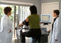
Cardiac/Cardiopulmonary Exercise Stress Testing
Exercise stress testing is used to assess a patient's
response of their heart, lungs and muscles to exercise.
The test obtains measurements related to the metabolic,
cardiovascular and pulmonary adaptations of the patient
from rest to maximum exercise and recovery. The exercise
test is performed by a technologist and a physician
experienced in exercise testing in a carefully controlled
setting. Every few minutes the elevation and speed
of the treadmill is increased and pedal resistance
is increased on the bike. Heart rate, electrocardiogram,
collecting and analyzing expired air, oxygen saturation
and blood pressure are monitored during cardiopulmonary
stress testing. The Bruce Protocol or Naughton Protocol
is used for basic Cardiac Assessment with the heart
rate, electrocardiogram and blood pressure being
monitored. The test continues until the patient reaches
his/her maximum exercise tolerance or pre-determined
safety limits have been exceeded.
Our Echocardiograph Stress Tests are preformed
on a treadmill, while the Cardiopulmonary exercise
stress test is performed on a bike.
These tests are very effective for patients who
are experiencing shortness of breath or chest pains.
The total time involved is 45 minutes to an hour.
Vascular
Investigations
Peripheral Doppler Testing:
The ankle brachial index (ABI) is a simple quick
comparison of blood pressure readings. This procedure
is similar to taking a blood pressure but a Doppler
is used instead of a stethoscope. The ABI is a non-invasive
procedure which will indicate common circulatory
problems in the extremities. The test involves placing
a blood pressure cuff on each arm and leg. Once the
measurements are taken the patient walks on the treadmill
for 5 minutes after which 3 pressures are taken.
Patients may have this test if they are unable to
walk a distance without experiencing pain. There
also could be pain experienced at rest. Other symptoms
include cyanosis, where the leg turns blue.
The total time involved is 45 minutes to an hour.
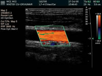
Carotid and Vertebral Doppler:
The Carotid Doppler is an ultrasound done on the
carotid arteries of the neck. This test is done to
check the blood flow of the arteries to observe if
there are any flow restrictions on either side.
Patients may have this test if they have had a previous
stroke. Other symptoms are dizziness, transient stroke,
or loss of vision. It can also be done on asymptomatic
patients pre/post operation, or with high risk factors.
The total time involved is 45 minutes to an hour.
Venous Colour Doppler:
The Venous Doppler is an ultrasound done on the
veins of the legs. This test is done to check if
there are any blood clots in the veins. The vein
will be found in the groin region and followed down
to the legs.
Patients will have this test if they experience
leg pain, swelling, tenderness, or discoloration.
The total time involved is 45 minutes to an hour.
|